Radiography
What is it?
Radiography is technique for imaging using X-rays which is used to visualize large areas of the body.
It helps to study the bones of the skeleton and joints, lungs, abdomen and breasts in order to help the doctor in the process of making a diagnosis.
Preparation
For your investigation, you must bring along the following:
- Your prescription
- Your previous radiological investigations for the concerned region of the body, if available
- Your insurance company contact details
Procedure
It is a very quick investigation (some seconds for each film of radiography, some minutes for the complete investigation of the body), non-invasive.
In case of painful pathology (fracture…), we are at your service and we will suggest solutions that are tailored to your needs.
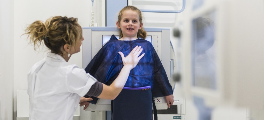
After you have worn a robe, we will position you – sitting, standing or lying down – depending on the region to be examined, following the prescriptions of your doctor.
A technician will capture the image(s) while asking you not to move during the duration of the capture, which is important to ensure images of good quality.
A lead apron may be used (if it does not hinder the investigation) to protect you from radiation.
Radiation
No risk has been demonstrated till the present time, considering the low doses used and the precautions taken by the technician to delimit the part of the body that is irradiated during each investigation.
An ordinary radiograph exposes you to the same quantity of X-rays as that you receive naturally from the sun during average flight duration of 4 hours.
However, at the Servette Imaging Centre, investigations must always be justified and precautions must be taken.
A radiographic examination does not require any particular examination (except for investigations of the gastrointestinal tract).
Pregnancy is a relative contraindication to radiography; a discussion must take place between the treating doctor, radiologist and patient in order to assess the risk-benefit ratio.
Results
As from the following day, the diagnosis will be transmitted by fax or in the form of a detailed report to the doctor who ordered the investigation.
Afterwards, you will also be able to request for the report as well as the images for 10 years after your investigation.
Our Machines
We are equipped with a latest generation device: SIEMENS AXIOM LUMINOS Drf.
This 100% digital technology has a flat panel on which radiographs can be viewed instantly via wifi connection and allows us to reduce the dose of radiation both for the patient and the care provider.
The system brings these two techniques together: fluoroscopy and radiography, which makes it possible to perform all the investigations at the Servette Imaging Centre with efficiency, safety and patient comfort.
An orthopedic support is also available in the room which makes it possible for large field radiographs to be taken.
Investigations performed
The team of Servette Imaging Centre performs radiographs on any part of the body.
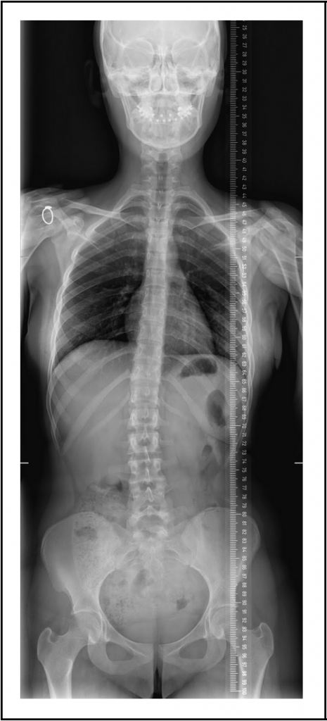
Full radiograph of the vertebral column
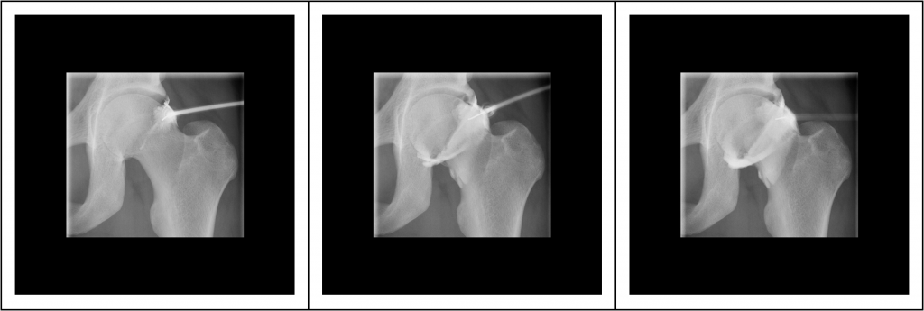
Arthrography of the hip joint
Upper gastrointestinal series
![]()
