What is it?
Mammography is radiography of the breast (using X-rays) which makes it possible to visualise very small benign or malignant tumours before they become palpable or manifest with other symptoms. It is also performed on breasts that have implants filled with silicone or saline solution.
In the absence of a family history or particular breast symptoms, this investigation is recommended every 2 years beginning from age 50 and may be requested either by your gynaecologist or by the cantonal screening programme.
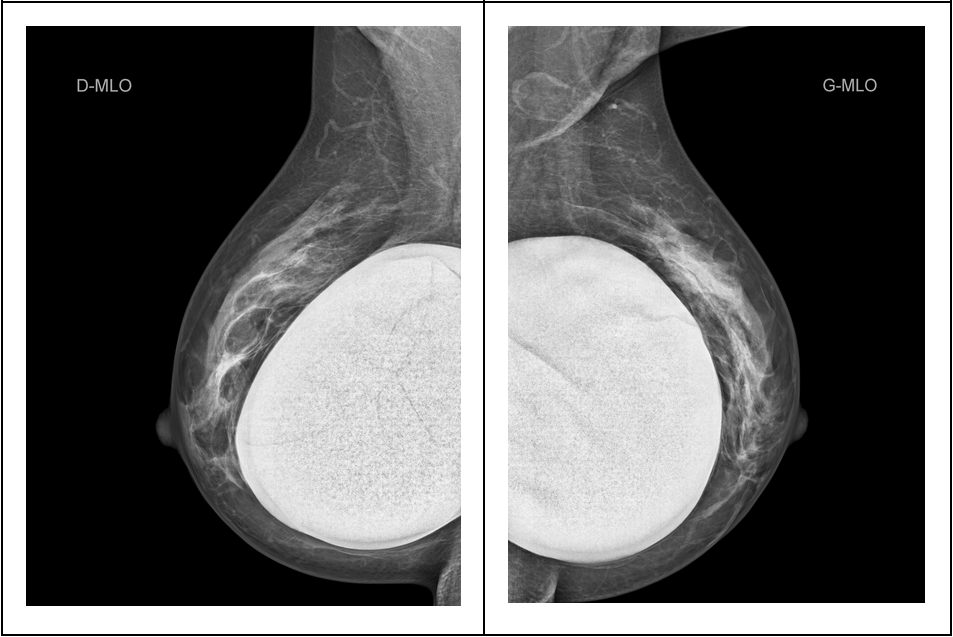
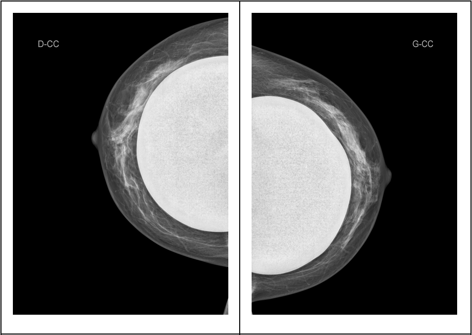
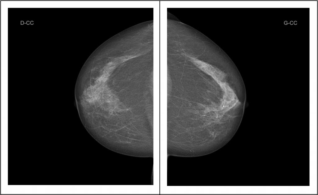
For a complete investigation, it is necessary to obtain 2 radiographs in different projections on each breast, one from the front and the other from the side, for a total of 4 images. In order to ensure a high quality of the image, the breast must be kept compressed during the emission of X-rays (10 seconds). This compression makes it possible to prevent motion blurs that may arise due to movement, to achieve better differentiation of the various structures of the mammary gland and to reduce the dose of radiation required.
Mammography must be performed between the 5th and 12th day following the 1st day of the menstrual cycle for pre-menopausal patients. This is in order to avoid possible unknown early pregnancy, reduce the discomfort during compression which is due to breast pressure during the premenstrual phase and to exclude benign breast pathologies which may occasionally appear during the rest of the cycle and hinder the visualisation of the mammary gland.
Preparation
You must bring along the following for your investigation:
- Your prescription
- Your past breast examination reports
- The contact details of your insurance company
Ensure that you are in the right period of your menstrual cycle (between day 5 and day 12 of the menstrual cycle).
Procedure
The technician in charge of your mammography will help you to fill a questionnaire for the purpose of providing the radiologist with information on your personal and family history as well as your current treatments.
Once you have undressed (bare torso) in the investigation room, we will proceed to capture the images: while making sure you are comfortable, we will compress your breasts one after the order in a very gradual manner.
This rather popular investigation is not painful if the procedure is gentle and well-explained.
At the Versoix Imaging Centre, we are convinced that a trust-based relationship between the technician and the patient is the key to a successful and well-tolerated investigation.
It should be remembered that the very brief compression is necessary in order to provide high-quality images and to reduce the dose of radiation applied to the breast (only 10 seconds).
A lead guard for the thyroid gland may be used if you request for it, but since the radiation is low and focused on the breast, the guard will be more of a disturbance than protection to you.
Results
After the investigation, we will usually take you to the ultrasonography room where the radiologist will perform an ultrasound scan.
Subsequently, the technician will give you an internet access which will make it possible for you to visualise your images and read the medical report.
As from the following day, the diagnosis will be transmitted to your prescribing physician, who will receive a detailed report by fax or telephone.
In future, you will also be able to request for the report as well as the images for up to 10 years after your investigation.
Our Machines
Our PHILIPS Microdose mammography machines provide excellent detection effectiveness while making use of very low doses.
In order to make the radiologist’s work and performance easier, image analysis is performed on high resolution computer screens.
Investigations performed
Investigations that complement mammography are essentially the following:
- Breast ultrasound
- One or several zoomed images of an area of the breast (during compression)
- Biopsy under ultrasound guidance: sampling of breast tissue using a needle
- Breast MRI: will be available soon.
Classic mammography

Mammography for silicone implants